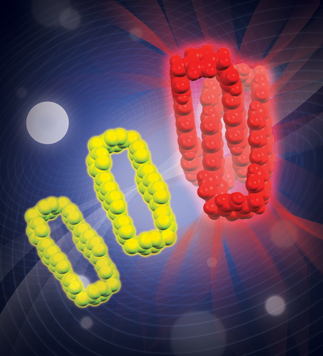
Dr Nancy McGuire writes in a JACS Spotlight that the fluorescent tagging of molecules have become mainstays for imaging biological systems. Watching where these tags attach and what conditions make them light up provides insights into cellular structures and processes. So-called excimers and exciplexes, aggregates of tagging molecules that emit brief bursts of low-energy light, have been considered a nuisance that reduces the fluorescence efficiency of single-molecule tags. However, Amine Garci and co-authors recently reported a family of mechanically interlocked molecules (MIMs) emitting long-lived and well-controlled exciplex fluorescence, even at the very dilute concentrations needed for imaging living cells (DOI: 10.1021/jacs.0c02128).
Their MIMs—stacked pairs of linked, elongated oval molecules—contain anthracene units built into a cyclophane framework. The anthracene acts as a self-templating agent that helps the aggregates form. It also holds the light-generating parts of the molecules close enough together to keep the exciplex fluorescence going. The authors imaged live prostate cancer cells using their MIMs, and they detected fluorescent red or green light emissions (depending on the type of tag) even at micromolar concentrations. Because these MIMs were readily admitted into the cells, only a small amount was needed to produce a detectable signal. This ease of cellular access could also make these MIMs useful as drug delivery agents.


